
Actinic keratosis (also called "solar keratosis" and "senile keratosis"; abbreviated as "AK") is a pre-cancerous patch of thick, scaly, or crusty skin. These growths are more common in fair-skinned people and those who are frequently in the sun. They usually form when skin gets damaged by ultraviolet (UV) radiation from the sun or indoor tanning beds. AKs are considered potentially pre-cancerous; left untreated, they may turn into a type of cancer called squamous cell carcinoma. Untreated lesions have up to a 20% risk of progression to squamous cell carcinoma, so treatment by a dermatologist is recommended.
Development of these growths occur when skin is constantly exposed to the sun over time. They usually appear as thick, scaly, or crusty areas that often feel dry or rough. In fact, AKs are often felt before they are seen, and the texture is often compared to sandpaper. They may be dark, light, tan, pink, red, a combination of all these, or have the same color as the surrounding skin. An actinic keratosis lesion commonly ranges between 2 and 6 millimeters in size but can grow to be a few centimeters in diameter. They often appear on sun-exposed areas of the skin, such as the face, ears, neck, scalp, chest, backs of hands, forearms, or lips. Because they are related to sun-damage on the skin, most people who have an AK have more than one.
Diagnosis is made clinically on physical exam, but can be confirmed by looking at cells from the lesion under a microscope. There are various options for treatment, but 5-Fluorouracil cream seems to be popular and effective. By following up with a dermatologist, AKs can be treated before they turn into skin cancer. If skin cancer does develop from an AK lesion, it can be caught early with close monitoring, at a time when treatment can be curative.
Signs and symptoms

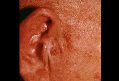
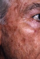
Actinic keratoses ("AKs") most commonly present as a white, scaly plaque of variable thickness with surrounding redness; they are most notable for having a sandpaper-like texture when felt with a gloved hand. Skin nearby the lesion often shows evidence of solar damage characterized by notable pigmentary alterations, being yellow or pale in color with areas of hyperpigmentation; deep wrinkles, coarse texture, purpura and ecchymoses, dry skin, and scattered telangiectasias are also characteristic. Photoaging leads to an accumulation of oncogenic changes, resulting in a proliferation of mutated keratinocytes that can manifest as AKs or other neoplastic growths. With years of sun damage, it is possible to develop multiple AKs in a single area on the skin. The lesions are usually asymptomatic, but can be tender, itch, bleed, or produce a stinging or burning sensation. AKs are typically graded in accordance with their clinical presentation: Grade I (easily visible, slightly palpable), Grade II (easily visible, palpable), and Grade III (frankly visible and hyperkeratotic).
Clinical variants
Actinic keratoses can have various clinical presentations, often characterized as follows:
- Classic (or common): Classic AKs present as white, scaly macules, papules or plaques of various thickness, often with surrounding erythema. They are usually 2-6mm in diameter but can sometimes reach several centimeters in diameter.
- Hypertrophic (or hyperkeratotic): Hypertrophic AKs (HAKs) appear as a thicker scale or rough papule or plaque, often adherent to an erythematous base. Classic AKs can progress to become HAKs, and HAKs themselves can be difficult to distinguish from malignant lesions.
- Atrophic: Atrophic AKs lack an overlying scale, and therefore appear as a nonpalpable change in color (or macule). They are often smooth and red, and are less than 10mm in diameter.
- AK with cutaneous horn: A cutaneous horn is a keratinic projection with its height at least one-half of its diameter, often conical in shape. They can be seen in the setting of actinic keratosis as a progression of an HAK, but are also present in other skin conditions. 38-40% of cutaneous horns represent AKs.
- Pigmented AK: Pigmented AKs are rare variants that often present as macules or plaques that are tan to brown in color. They can be difficult to distinguish from a solar lengtigo or lentigo maligna.
- Actinic cheilitis: When an AK forms on the lip, it is called actinic cheilitis. This usually presents as a rough, scaly patch on the lip, often accompanied by the sensation of dry mouth and symptomatic splitting of the lips.
- Bowenoid AK: Usually presents as a solitary, erythematous, scaly patch or plaque with well-defined borders. Bowenoid AKs are differentiated from Bowen's disease by degree of epithelial involvement as seen on histology.
The presence of ulceration, nodularity, or bleeding should raise concern for malignancy. Specifically, clinical findings suggesting an increased risk of progression to squamous cell carcinoma can be recognized as:Â IDRBEU. I (Induration /Inflammation), D (Diameter > 1Â cm), R (Rapid Enlargement), B (Bleeding), E (Erythema) and U (Ulceration). AKs are usually diagnosed clinically, but because they are difficult to clinically differentiate from squamous cell carcinoma, any concerning features warrant biopsy for diagnostic confirmation.
Clinical course
Actinic keratoses have three possible clinical outcomes: they may regress, remain stable, or advance to become invasive disease. Occasionally they come and go, appearing on the skin, remaining for months, and then disappearing. Often they will reappear in a few weeks or months, particularly after unprotected sun exposure. Left untreated, there is a chance that the lesion will advance to become invasive. While it is difficult to predict whether an AK will advance to become squamous cell carcinoma, it has been noted that squamous cell carcinomas originate in lesions formerly diagnosed as AKs with frequencies reported between 65 to 97%.
Cause

The most important cause of AK formation is solar radiation, specifically UV-B radiation (wavelength 290-320). UV-B radiation causes thymidine dimer formation in DNA and RNA, leading to significant cellular mutations. Additionally, recent research has been focused on the role of the p53 tumor suppressor gene in the role of AK formation. This tumor suppressor gene, located on chromosome 17p132 allows for cell cycle arrest when DNA or RNA is damaged. Dysregulation of the p53 pathway can thus result in unchecked proliferation of dysplastic keratinocytes, thereby serving as a source of neoplastic growth and the development of AK, as well as possible carcinogenesis. Other molecular markers that have been associated with the development of AK include the expression of p16ink4, the CD95 ligand, TNF-related apoptosis-inducing ligand (TRAIL) and TRAIL receptors, and loss of heterozygosity.
Recent research suggests that the human papillomavirus (HPV) may also play a role in the development of AKs. The HPV virus has been detected in AKs, with measurable HPV viral loads (1 HPV-DNA copy per less than 50 cells) measured in 40% of AKs.Similar to UV radiation, higher levels of HPV found in AKs reflect enhanced viral DNA replication; this is suspected to be related to the abnormal keratinocyte proliferation and differentiation in AKs which provide a commensalic environment for HPV replication. This in turn may further stimulate the abnormal proliferation that contributes to the development of AKs and carcinogenesis.
AKs are most often seen in individuals with fair skin and are commonly found on the scalp of balding individuals.
Ultraviolet radiation
It is thought that UV radiation induces mutations in the keratinocytes of the epidermis, and promoting both the survival proliferation of these atypical cells. Eventually, this leads to the formation of AKs. In particular, mutations in the p53 tumor suppressor gene have been found in 30-50% of AK lesion skin samples.
- Extent of sun exposure: Cumulative sun exposure leads to an increased risk for development of AKs. In one U.S. study, AKs were found in 55% of fair-skinned men with high cumulative sun exposure, and in only 19% of fair-skinned men with low cumulative sun exposure in an age-matched cohort (the percents for women in this same study were 37% and 12% respectively). Furthermore, the use of sunscreen (SPF 17 or higher) has been found to significantly reduce the development of AK lesions, and also promotes the regression of existing lesions.
- History of sunburn: Studies show that even a single episode of painful sunburn as a child can increase an individual's risk of developing AK as an adult. Six or more painful sunburns over the course of a lifetime was found to be significantly associated with the likelihood of developing AK.
Skin pigmentation
Melanin is a pigment in the epidermis that functions to protect keratinocytes from the damage caused UV radiation; it is found in higher concentration in the epidermis of darker-skinned individuals, affording them protection against the development of AKs. Fair-skinned individuals have a significantly increased risk of developing AKs when compared to olive skinned individuals (odds ratios of 14.1 and 6.5, respectively), and AKs are uncommon in dark-skinned African Americans. Other phenotypic features seen in fair-skinned individuals that are associated with an increased propensity to develop AKs include:
- Freckling
- Light hair color
- Propensity to sunburn
- Inability to tan
Balding
AKs are not uncommonly found on the scalps of balding men. Degree of baldness seems to be a risk factor for lesion development, as men with severe baldness were found to be seven times more likely to have 10 or more AKs when compared to men with minimal or no baldness.
Other risk factors
- Immunosuppression: People who take immunosuppressive drugs, such as organ transplant patients, are 250 times more likely to develop actinic keratoses that may lead to skin cancer.
- Human papillomavirus (HPV): The role of HPV in the development of AK remains unclear, but evidence suggests that infection with the betapapillomavirus type of HPV may be associated with an increased likelihood of AK.
- Genodermatoses: Certain genetic disorders interfere with DNA repair after sun exposure, thereby putting these individuals at higher risk for the development of AKs. Examples of such genetic disorders include xeroderma pigmentosum and Bloom syndrome.
Diagnosis
Physicians usually diagnose actinic keratosis by doing a thorough physical examination, through a combination of visual observation and touch. However a biopsy may be necessary when the keratosis is large in diameter, thick, or bleeding, in order to make sure that the lesion is not a skin cancer. Actinic keratosis and squamous cell carcinoma (SCC) can present similarly on physical exam, and many scientists argue that they are in fact simply different stages of the same condition. In addition to SCCs, AKs can be mistaken for other cutaneous lesions including: seborrheic keratoses, basal cell carcinoma, lichenoid keratosis, porokeratosis, viral warts, inflammatory dermatoses, or melanoma.
Biopsy
A lesion biospy is performed if the diagnosis remains uncertain after a clinical physical exam. The most common tissue sampling techniques include shave or punch biopsy. When only a portion of the lesion can be removed due to its size or location, the biopsy should sample tissue from the thickest area of the lesion, as SCCs are most likely to be detected in that area. If a shave biopsy is performed, it should extend through to the level of the dermis in order to provide sufficient tissue for diagnosis; ideally, it would extend to the mid-reticular dermis. Punch biopsy usually extends to the subcutaneous fat when the entire length of the punch blade is utilized.
Histopathology
On histologic examination, actinic keratoses usually show a collection of atypical keratinocytes with hyperpigmented or pleomorphic nuclei, extending to the basal layer of the epidermis. A "flag sign" is often described, referring to alternating areas of orthokeratosis and parakeratosis. Epidermal thickening and surrounding areas of sun-damaged skin are often seen. The normal ordered maturation of the keratinocytes is disordered to varying degrees: there may be widening of the intracellular spaces, cytologic atypia such as abnormally large nuclei, and a mild chronic inflammatory infiltrate.
Specific findings depend on the clinical variant and particular lesion characteristics. The seven major histopathologic variants are all characterized by atypical keratinocytic proliferation beginning in the basal layer and confined to the epidermis; they include:
- Hypertrophic: Notable for marked hyperkeratosis, often with evident parakeratosis. Keratinocytes in the stratum malphigii may show a loss of polarity, pleomorphism, and anaplasia. Some irregular downward proliferation into the uppermost dermis may be observed, but does not represent frank invasion.
- Atrophic: With slight hyperkeratosis and overall atrophic changes to the epidermis; the basal layer shows cells with large, hyperchromatic nuclei in close proximity to each other. These cells have been observed to proliferate into the dermis as buds and duct-like structures.
- Lichenoid: Demonstrate a band-like lymphocytic infiltrate in the papillary dermis, directly beneath the dermal-epidermal junction.
- Achantholytic: Intercelleular clefts or lacunae in the lowermost epidermal layer that result from anaplastic changes; these produce dyskeratotic cells with disrupted intercellular bridges.
- Bowenoid: This term is controversial and usually refers to full-thickness atypia, microscopically indistinguishable from Bowen's Disease. However most dermatologists and pathologists will use it in reference to tissue samples that are notable for small foci of atypia that involve the full thickness of the epidermis, in the the background of a lesion that is otherwise consistent with an AK.
- Epidermolytic: With granular degeneration.
- Pigmented: Show pigmentation in the basal layer of the epidermis, similar to a solar lentigo.
Dermoscopy
Dermoscopy is a noninvasive technique utilizing a handheld magnifying device coupled with a transilluminating lift. It is often used in the evaluation of cutaneous lesions, but lacks the definitive diagnostic ability of biopsy-based tissue diagnosis. Histopathologic exam remains the gold standard
Dermoscopic features
Polarized contact dermoscopy of AKs occasionally reveals a "rosette sign," described as four white points arranged in a clover pattern, often localized to within a follicular opening. It is hypothesized that the "rosette sign" corresponds histologically to the changes of orthokeratosis and parakeratosis known as the "flag sign."
- Non-pigmented AKs: linear or wavy vascular patterning, or a "strawberry pattern," described as unfocused vessels between hair follicles, with white-haloed follicular openings.
- Pigmented AKs: gray to brown dots or globules surrounding follicular openings, and annular-granular rhomboidal structures; often difficult to differentiate from lentigo maligna.
Prevention
Ultraviolet radiation is believed to contribute to the development of actinic keratoses (AKs) by inducing mutations in epidermal keratinocytes, leading to proliferation of atypical cells. Therefore, preventive measures for AKs are targeted at limiting exposure to solar radiation, including:
- Limiting extent of sun exposure
- Avoid sun exposure during noontime hours when UV light is most powerful
- Using sun protection
- Frequently applying powerful sunscreens with SPF ratings greater than 30 and that also block both UVA and UVB light
- Wearing sun protective clothing such as hats, long-sleeved shirts, long skirts, or trousers
Recent research implicating human papillomavirus (HPV) in the development of AKs suggest that HPV prevention might in turn help prevent development of AKs, as UV-induced mutations and oncogenic transformation are likely facilitated in cases of active HPV infection.
Management
There are many treatment options for AK depending on the patient and the clinical characteristics of the lesion. AKs show a wide range of features, which guide treatment decision-making. Although overall cure rates are high, experts agree that the best treatment for AK is prevention. Regular follow-up is advisable after any treatment to make sure no new lesions have developed and that old ones are not progressing.
Medication
Fluorouracil cream
Topical fluorouracil (5-FU) destroys AKs by blocking methylation of thymidylate synthetase, thereby interrupting DNA and RNA synthesis. This in turn prevents the proliferation of dysplastic cells in AK. Topical 5-FU is the most utilized treatment for AK, and often results in effective removal of the lesion. Overall, there is a 50% efficacy rate resulting in 100% clearance of AKs treated with topical 5-FU. 5-FU may be up to 90% effective in treating non-hyperkeratotic lesions. The most commonly used application regimen consists of applying a layer of topical cream to the lesion twice a day after washing; duration of treatment is typically 2â€"4 weeks, but treatment of up to 8 weeks has demonstrated a higher cure rate.
Imiquod cream
Imiquod is a topical immune-enhancing agent licensed for the treatment of genital warts. Imiquod stimulates the immune system through the release and up-regulation of cytokines. Treatment with imiquod cream applied 2-3 times per week for 12 to 16 weeks was found to result in complete resolution of AKs in 50% of people, compared to 5% of controls. The imiquod 3.75% cream has been validated in a treatment regimen consisting of daily application to entire face and scalp for two 2-week treatment cycles, with a complete clearance rate of 36%. While the clearance rate observed with the imiquod 3.75% cream was lower than that observed with the 5% cream (36 and 50 percent, respectively), there are lower reported rates of adverse reactions with the 3.75% cream: 19% of individuals using imiquod 3.75% cream reported adverse reactions including local erythema, scabbing, and flaking at the application site, while nearly a third of individuals using the 5% cream reported the same types of reactions with imiquod treatment. However it is ultimately difficult to compare the efficacy of the different strength creams directly, as current study data varies in methodology (e.g. duration and frequency of treatment, and amount of skin surface area covered).
Ingenol mebutate gel
Ingenol mebutate is a newer treatment for AK used in Europe and the United States. It works in two ways, first by disrupting cell membranes and mitochondria resulting cell death, and then by inducing antibody-dependent cellular cytotoxicity to eliminate remaining tumor cells. A 2-day treatment course with the 0.015% gel is recommended for the scalp and face, while a 3-day treatment course with the 0.05% gel is recommended for the trunk and extremities. Treatment with the 0.015% gel was found to completely clear 57% of AK, while the 0.05% gel had a 34% clearance rate. Advantages of ingenol mebutate treatment include the short duration of therapy and a low recurrence rate. Local skin reactions including pain, itching and redness can be expected during treatment with ingenol mebutate.
Diclofenac sodium gel
Topical diclofenac sodium gel is a nonsteroidal anti-inflammatory drug that is thought to work in the treatment of AK through its inhibition of the arachidonic acid pathway, thereby limiting the production of prostaglandins which are thought to be involved in the development of UVB-induced skin cancers. Recommended duration of therapy is 60 to 90 days with twice daily application. Treatment of facial AK with diclofenac gel led to complete lesion resolution in 40% of cases. Common side effects include dryness, itching, redness, and rash at the site of application.
Retinoids
Topical retinoids have been studied in the treatment of AK with modest results. Treatment with adapalene gel daily for 4 weeks, and then twice daily thereafter for a total of nine months led to a significant but modest reduction in the number AKs compared to placebo; it demonstrated the additional advantage of improving the appearance of photodamaged skin. Topical tretinoin is ineffective as treatment for reducing the number of AKs. For secondary prevention of AK, systemic, low dose aciretin was found to be safe, well-tolerated and moderately effective in chemoprophylaxis for skin cancers in renal transplant patients.
Procedures
Cryotherapy
Liquid nitrogen (âˆ'195.8 °C) is the most commonly used destructive therapy for the treatment of AK. It is a well-tolerated office procedure that does not require anesthesia. Cryotherapy is particularly indicated for cases where there are few, thin, well-demarcated lesions. It is generally performed using an open-spray technique, wherein the AK is sprayed for several seconds. The process can be repeated multiple times in one office visit, as tolerated. Cure rates from 67 to 99 percent have been reported, depending on freeze time and lesion characteristics. Disadvantages include discomfort during and after the procedure; blistering, scarring and redness; hypo- or hyper pigmentation; and destruction of healthy tissue.
Photodynamic therapy
AKs are one of the most common dermatologic lesion for which Photodynamic therapy using topical methyl aminolevulinate (MAL) or 5-aminolevulinic acid (5-ALA) is indicated. Treatment begins with preparation of the lesion, which includes scraping away scales and crusts using a dermal curette. A thick layer of topical MAL or 5-ALA cream is applied to the lesion and a small area surrounding the lesion, which is then covered with an occlusive dressing and left for a period of time. During this time the photosensitized accumulates in the target cells within the AK lesion. The dressings are then removed and the lesion is treated with light at a specified wavelength. Mulitple treatment regimens using different photosensitizers, incubation times, light sources, and pretreatment regimens have been studied and suggest that longer incubation times lead to higher rates of lesion clearance. Photodynamic therapy is gaining in popularity. It has been found to have a 14% higher likelihood of achieving complete lesion clearance at 3 months compared to cryotherapy, and seems to result in superior cosmetic outcomes when compared to cryotherapy or 5-FU treatment. Photodynamic therapy seems particularly effective in treating areas with multiple AK lesions.
Surgical techniques
- Surgical excision: Excision should be reserved for cases when the AK is a thick, horny papule, or when deeper invasion is suspected and histopathologic diagnosis is necessary. It is a rarely utilized technique for AK treatment.
- Shave excision and curretage (frequently followed by electrodessication): This technique is often used for treatment of AKs, and particularly for hyperkeratotic lesions. The surface of the lesion can be scraped away using a scalpel, or the base can be removed with a curette. Tissue can be evaluated histopathologically, but specimens acquired using this technique are not often adequate to determine whether a lesion is invasive or intraepidermal.
- Dermabrasion: Dermabrasion is useful in the treatment of large areas with multiple AK lesions. The process involves using a hand-held instrument to "sand" the skin, removing the stratum corneum layer of the epidermis. Diamond fraises or wire brushes revolving at high speeds are used. The procedure can be quite painful and requires procedural sedation and anesthetic, necessitating a hospital stay. One-year clearance rates with dermabrasion treatment are as high as 96%, but diminish drastically to 54% at five years.
Laser therapy
Laser therapy using carbon dioxide (CO
2) or erbium:yttrium aluminum garnet (Er:YAG) lasers is a treatment approach being utilized with increased frequency, and sometimes in conjunction with computer scanning technology. Laser therapy has not been extensively studied, but current evidence suggests it may be effective in cases involving multiple AKs refractive to medical therapy, or AKs located in cosmetically relevant locations such as the face.
Chemical peels
A chemical peel is a topically applied agent that wounds the outermost layer of the skin, promoting organized repair, exfoliation, and eventually the development of smooth and rejuvenated skin. Multiple therapies have been studied. A medium-depth peel may effectively treat multiple non-hyperkeratotic AKs. It can be achieved with 35% to 50% trichloroacetic acid (TCA) alone or at 35% in combination with Jessner's solution in a once-daily application for a minimum of 3 weeks; 70% glycolic acid (α-hydroxy acid); or solid CO
2. When compared to treatment with 5-FU, chemical peels have demonstrated similar efficacy and increased ease of use with similar morbidity. Chemical peels must be performed in a controlled clinic environment and are only recommended for individuals who are able to comply with follow-up precautions, including avoidance of sun exposure. Furthermore, they should be avoided in individuals with a history of HSV infection or keloids, and in those who are immunosuppressed or who are taking photosensitizing medications.
Prognosis

AKs follow one of three paths: they can either persist as AKs, regress, or progress to invasive skin cancer, as AKs lesions are considered to be on the same continuum with squamous cell carcinoma (SCC). AK lesions that regress also have the potential to recur.
- Progression: The overall risk of an AK turning into invasive cancer is low. In average-risk individuals, likelihood of an AK lesion progressing to SCC is less than 1% per year. Despite this low rate of progression, studies suggest that a full 60% of SCCs arise from pre-existing AKs, reinforcing the idea that these lesions are closely related.
- Regression: Reported regression rates for single AK lesions have ranged between 15-63% after one year.
- Recurrence: Recurrence rates after 1 year for single AK lesions that have regressed range between 15-53%.
Epidemiology
Actinic keratosis is very common, with an estimated 14% of dermatology visits related to AKs. It is seen more often in fair-skinned individuals, and rates vary with geographical location and age. Other factors such as exposure to ultraviolet (UV) radiation, certain phenotypic features, and immunosuppression can also contribute to the development of AKs.
Men are more likely to develop AK than women, and the risk of developing AK lesions increases with age. These findings have been observed in multiple studies, with numbers from one study suggesting that approximately 5% of women ages 20-29 develop AK compared to 68% of women ages 60-69, and 10% of men ages 20-29 develop AK compared to 79% of men ages 60-69.
Geography seems to play a role in the sense that individuals living in locations where they are exposed to more UV radiation throughout their lifetime have a significantly higher risk of developing AK. Much of the literature on AK comes from Australia, where prevalence of AK is estimated at 40-50% in adults over 40, as compared to the United States and Europe, where prevalence is estimated at under 11-38% in adults. One study found that those who immigrated to Australia after age 20 had fewer AKs than native Australians in all age groups.
Research

Diagnostically, researchers are investigating the role of novel biomarkers to assist in determining which AKs are more likely to develop into cutaneous or metastatic SCC. Upregulation of matrix metalloproteinases (MMP) is seen in many different types of cancers, and the expression and production of MMP-7 in particular has been found to be elevated in SCC specifically. The role of serin peptidase inhibitors (Serpins) is also being investigated. SerpinA1 was found to be elevated in the keratinocytes of SCC cell lines, and SerpinA1 upregulation was correlated with SCC tumor progression in vivo. Further investigation into specific biomarkers could help providers better assess prognosis and determine best treatment approaches for particular lesions.
In terms of treatment, a number of medications are being studied. Resiquimod is a TLR 7/8 agonist that works similarly to imiquimod, but is 10 to 100 times more potent; when used to treat AK lesions, complete response rates have range from 40 to 74%. Afamelanotide is drug that induces the production of melanin by melanocytes to act as a protective factor against UVB radiation. It is being studied to determine its efficacy in preventing AKs in organ transplant patients who are on immunosuppressive therapy. Epidermal growth factor receptor (EGFR) inhibitors such as gefitinib, and anti-EGFR antibodies such as cetuximab are used in the treatment of various types of cancers, and are currently being investigated for potential use in the treatment and prevention of AKs.
References
External links
- Actinic Keratosis at Mediconum.com
- American Academy of Dermatology
- Actinic Keratosis photo library at Dermnet