
Amblyopia (from Greek αμβλυωπία, "blunt vision"), also called lazy eye, is a disorder of sight. It involves decreased vision in an eye that otherwise appears normal, or out of proportion to associated structural problems of the eye; there is much more 'damage to' or impact on vision in that eye than is predicted, even though the eye doesn't look completely normal. This disorder has been estimated to affect 1-5% of the population.
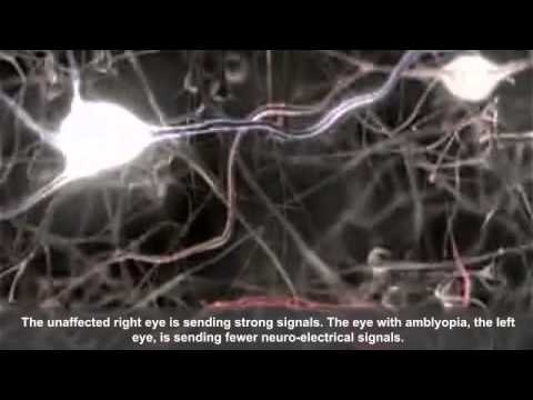
In amblyopia, visual stimulation either fails to be or is poorly transmitted through the optic nerve to the brain for a continuous period of time. It can also occur when the brain "turns off" the visual processing of one eye to prevent double-vision, for example in strabismus (crossed eyes). It often occurs during early childhood and results in poor or blurry vision.
Detecting the condition in early childhood increases the chance of successful treatment, especially if detected before the age of five. The earlier it is detected, and the underlying cause corrected with glasses or surgery, the better the long term outcomes.
Signs and symptoms
Many people with amblyopia, especially those who only have a mild form, are not even aware they have the condition until tested at older ages, since the vision in their stronger eye is normal. However, people who have severe amblyopia may experience related visual disorders, most notably poor depth perception. Amblyopes may suffer from poor spatial acuity, low sensitivity to contrast motion. Amblyopia is characterized by several functional abnormalities in spatial vision, including reductions in visual acuity (VA), contrast sensitivity function (CSF), and vernier acuity as well as spatial distortion, abnormal spatial interactions, and impaired contour detection. In addition, amblyopic individuals suffer from binocular abnormalities such as impaired stereoacuity (stereoscopic acuity) and abnormal binocular summation. Also, a crowding phenomenon is present. These deficits are usually specific to the amblyopic eye. However, sub-clinical deficits of the "better" eye have also been demonstrated.
People with amblyopia also have problems of binocular vision such as limited stereoscopic depth perception and usually have difficulty seeing the three-dimensional images in hidden stereoscopic displays such as autostereograms. However, perception of depth from monocular cues such as size, perspective, and motion parallax remains normal.
Types
Amblyopia has three main causes:
- Strabismic: by strabismus (misaligned eyes)
- Refractive: by anisometropia (high degrees of nearsightedness, farsightedness, or astigmatism in one or both eyes)
- Deprivational: by deprivation of vision early in life by vision-obstructing disorders such as congenital cataract
Strabismus amblyopia
Strabismus, sometimes also incorrectly called lazy eye, is a condition in which the eyes are misaligned. Strabismus usually results in normal vision in the preferred sighting (or "fellow") eye (the eye that the person prefers to use), but may cause abnormal vision in the deviating or strabismus eye due to the difference between the images projecting to the brain from the two eyes. Adult-onset strabismus usually causes double vision (diplopia), since the two eyes are not fixed on the same object. Children's brains, however, are more neuroplastic, and therefore can more easily adapt by suppressing images from one of the eyes, eliminating the double vision. This plastic response of the brain, however, interrupts the brain's normal development, resulting in the amblyopia. Recent evidence points to a cause of infantile strabism lying with the input to the visual cortex.
Strabismic amblyopes tend to show ocular motion deficits when reading, even when they use the nonamblyopic eye. In particular, they tend to make more saccades per line than persons with normal stereo vision, and to have a smaller reading speed, especially when reading a text with small font size.
Strabismus amblyopia is treated by clarifying the visual image with glasses, or encouraging use of the amblyopic eye with an eyepatch over the dominant eye or pharmacologic penalization of the better eye. Penalization usually consists of applying atropine drops to temporarily dilate the pupil, which leads to blurring of vision in the good eye. This helps to prevent the bullying and teasing associated with wearing a patch, although sometimes application of the eyedrops is more challenging. The ocular alignment itself may be treated with surgical or non-surgical methods, depending on the type and severity of the strabismus.
Refractive or anisometropic amblyopia
Refractive amblyopia may result from anisometropia (unequal refractive error between the two eyes). Anisometropia exists when there is a difference in the power between the two eyes. The eye which provides the brain with a clearer image typically becomes the dominant eye. The image in the other eye is blurred, which results in abnormal development of one half of the visual system. Refractive amblyopia is usually less severe than strabismic amblyopia and is commonly missed by primary care physicians because of its less dramatic appearance and lack of obvious physical manifestation, such as with strabismus. Given that the refractive correction of anisometropia by means of spectacles typically leads to different image magnification for the two eyes, which may in turn prevent binocular vision, a refractive correction using contact lenses is to be considered. Also pediatric refractive surgery is a treatment option, in particular if conventional approaches have failed due to aniseikonia or lack of compliance or both.
Frequently, amblyopia is associated with a combination of anisometropia and strabismus. In some cases, the vision between the eyes can differ to the point where one eye has twice average vision while the other eye is completely blind.
Deprivation and occlusion amblyopia
Deprivation amblyopia (Amblyopia ex anopsia) results when the ocular media become opaque, such as is the case with congenital cataract or corneal haziness. These opacities prevent adequate visual input from reaching the eye, and therefore disrupt development. If not treated in a timely fashion, amblyopia may persist even after the cause of the opacity is removed. Sometimes, drooping of the eyelid (ptosis) or some other problem causes the upper eyelid to physically occlude a child's vision, which may cause amblyopia quickly. Occlusion amblyopia may be a complication of a hemangioma that blocks some or all of the eye.
Pathophysiology

Amblyopia is a developmental problem in the brain, not any intrinsic, organic neurological problem in the eyeball (although organic problems can lead to amblyopia which can continue to exist after the organic problem has resolved by medical intervention). The part of the brain receiving images from the affected eye is not stimulated properly and does not develop to its full visual potential. This has been confirmed by direct brain examination. David H. Hubel and Torsten Wiesel won the Nobel Prize in Physiology or Medicine in 1981 for their work in showing the extent of the damage to ocular dominance columns produced in kittens by sufficient visual deprivation during the so-called "critical period." The maximum "critical period" in humans is from birth to two years old.
Treatment

Treatment of strabismic or anisometropic amblyopia consists of correcting the optical deficit (wearing the necessary spectacle prescription) and often forcing use of the amblyopic eye, by patching the good eye, or instilling topical atropine in the good eye, or both.
Concerning patching versus atropine, there is a drawback in using atropine: the drops can have a side effect of creating nodules in the eye which a correctional ointment can counteract. One should also be wary of over-patching or over-penalizing the good eye when treating for amblyopia, as this can create so-called "reverse amblyopia". Eye patching is usually done on a part-time schedule of about 4â€"6 hours a day. Treatment is continued as long as vision improves. It is not worthwhile continuing to patch for more than 6 months if there is no improvement.
Treatment of individuals age 9 through adult is possible through applied perceptual learning.
Deprivation amblyopia is treated by removing the opacity as soon as possible followed by patching or penalizing the good eye to encourage use of the amblyopic eye. The earlier treatment is initiated, the easier and faster the treatment is and the less psychologically damaging. There is also a greater chance of achieving 20/20 vision if treatment is initiated as early as possible.
One of the German public health insurance providers, Barmer, has changed its policy to cover, as of 1 April 2014, the costs for an app for amblyopic children whose condition has so far not improved through patching. The app offers dedicated eye exercises which the patient performs while wearing an eyepatch.
Older age
Although the best outcome is achieved if treatment is started before age 8, research has shown that children older than age 12 and some adults can show improvement in the affected eye. Children from 9 to 11 who wore an eye patch and performed near point activities (vision therapy) were four times as likely to show a two line improvement on a standard 11 line eye chart than amblyopic children who did not receive treatment. Adolescents aged 13 to 17 showed improvement as well, albeit in smaller amounts than younger children. It is uncertain whether such improvements are only temporary, however, particularly if treatment is discontinued.
There is tentative evidence that perceptual training may be beneficial in adults.
Virtual reality computer games where each eye receives different signals of the virtual world that the player's brain must combine in order to successfully play the game have shown some promise in improving both monocularity in the affected eye as well as binocularity.
Research

A study, widely reported in the popular press, has suggested that repetitive transcranial magnetic stimulation may temporarily improve contrast sensitivity and spatial resolution in the affected eye of amblyopic adults. This approach is still under development, and the results await verification by other researchers. It has also been suggested that comparable results can be achieved using different types of brain stimulation such as anodal transcranial direct current stimulation and theta burst rTMS.
The most recent study suggests that playing a version of the popular game Tetris that is modified such that each eye sees separate components of the game may also help to treat this condition in adults. Furthermore, it has been proposed that the effects of this kind of therapy may be further enhanced by non-invasive brain stimulation as shown by a recent study using anodal tDCS.
