The meninges (/məˈnɪndÊ'iËz/, singular: meninx (/ˈmiËnɪŋks/ or /ˈmÉ›nɪŋks/), from Ancient Greek: μῆνιγξ mÄ"ninx “membraneâ€, adjectival: meningeal /məˈnɪndÊ'É™l/) are the membranes that envelop the central nervous system. In mammals, the meninges consist of three layers: the dura mater, the arachnoid mater, and the pia mater. The primary function of the meninges and of the cerebrospinal fluid is to protect the central nervous system.
Structure

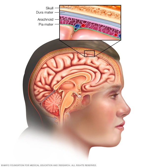
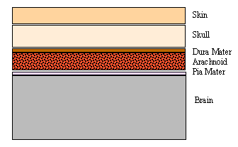
Dura mater
The dura mater [Latin: 'tough mother'] (also rarely called meninx fibrosa or pachymeninx) is a thick, durable membrane, closest to the skull. The dura mater, the outermost part, is a loosely arranged, fibroelastic layer of cells, characterized by multiple interdigitating cell processes, no extracellular collagen, and significant extracellular spaces. The middle region is a mostly fibrous portion. It consists of two layers: the endosteal layer, which lies closest to the calvaria (skull), and the inner meningeal layer, which lies closer to the brain. It contains larger blood vessels that split into the capillaries in the pia mater. It is composed of dense fibrous tissue, and its inner surface is covered by flattened cells like those present on the surfaces of the pia mater and arachnoid mater. The dura mater is a sac that envelops the arachnoid mater and surrounds and supports the large scrams channels (dural sinuses) carrying blood from the brain toward the heart.
The dura has four areas of infolding:
- Falx cerebri, the largest, sickle-shaped; separates the cerebral hemispheres. Starts from the frontal crest of frontal bone and the crista galli running to the internal occipital protuberance.
- Tentorium cerebelli, the second largest, crescent-shaped; separates the occipital lobes from cerebellum. The falx cerebri attaches to it giving a tentlike appearance.
- Falx cerebelli, vertical infolding; lies inferior to the tentorium cerebelli, separating the cerebellar hemispheres.
- Diaphragma sellae, smallest infolding; covers the pituitary gland and sella turcica.
Arachnoid mater
The middle element of the meninges is the arachnoid mater, so named because of its spider web-like appearance. It cushions the central nervous system. This thin, transparent membrane is composed of fibrous tissue and, like the pia mater, is covered by flat cells also thought to be impermeable to fluid.
The shape of the arachnoid does not follow the convolutions of the surface of the brain and so looks like a loosely fitting sac. In particular, in the region of the brain a large number of fine filaments called arachnoid trabeculae pass from the arachnoid through the subarachnoid space to blend with the tissue of the pia mater. The arachnoid is composed of an outermost portion (arachnoid barrier cell layer) with tightly packed cells and no extracellular collagen; that is why it is considered to represent an effective morphological and physiological meningeal barrier between the cerebrospinal fluid and subarachnoid space and the blood circulation in the dura.
The arachnoid barrier layer is characterized by a distinct continuous basal lamina on its inner surface toward the innermost collagenous portion of the arachnoid reticular layer.
Pia mater
The pia mater [Latin: 'soft mother'] is a very delicate membrane. It is the meningeal envelope that firmly adheres to the surface of the brain and spinal cord, following the brain's minor contours (gyri and sulci). It is a very thin membrane composed of fibrous tissue covered on its outer surface by a sheet of flat cells thought to be impermeable to fluid. The pia mater is pierced by blood vessels to the brain and spinal cord, and its capillaries nourish the brain.
Leptomeninges
The arachnoid and pia mater together, are sometimes called the leptomeninges, literally thin meninges. The morphology of the brain in meningococcal meningitis is said to be covered by exudate on the surface of and within the leptomeninges. Because the arachnoid is connected to the pia by cob-web like strands, it is structurally continuous with the pia, hence the name pia-arachnoid or leptomeninges.
Spaces
The subarachnoid space is the space that normally exists between the arachnoid and the pia mater, which is filled with cerebrospinal fluid.
Normally, the dura mater is attached to the skull, in the spinal cord, the dura mater is separated from the bone (vertebrae) by a space called the epidural space, which contain fat and blood vessels. The arachnoid is attached to the dura mater, while the pia mater is attached to the central nervous system tissue. When the dura mater and the arachnoid separate through injury or illness, the space between them is the subdural space. There is a subpial space underneath the pia mater that separates it from the glia limitans.
Clinical significance

There are three types of hemorrhage involving the meninges:
- A subarachnoid hemorrhage is acute bleeding under the arachnoid; it may occur spontaneously or as a result of trauma.
- A subdural hematoma is a hematoma (collection of blood) located in a separation of the arachnoid from the dura mater. The small veins that connect the dura mater and the arachnoid are torn, usually during an accident, and blood leaks into this area.
- An epidural hematoma may arise after an accident or spontaneously.
Other medical conditions that affect the meninges include meningitis (usually from fungal, bacterial, or viral infection) and meningiomas that arise from the meninges, or from meningeal carcinomatoses (tumors) that form elsewhere in the body and metastasize to the meninges.
In other animals
In fish, there is a single membrane (the primitive meninx). In amphibians, reptiles and birds, the meninges include a thick outer dura mater and a thick inner secondary meninx. Mammals retain the dura mater, and the secondary meninx divides into the arachnoid and pia mater.
Additional images

References