Ascaris lumbricoides is the giant roundworm of humans, growing to a length of up to 35Â cm. It is one of several species of Ascaris. An ascarid nematode of the phylum Nematoda, it is the largest and most common parasitic worm in humans. This organism is responsible for the disease ascariasis, a type of helminthiasis and one of the group of neglected tropical diseases. An estimated one-sixth of the human population is infected by A. lumbricoides or another roundworm. Ascariasis is prevalent worldwide, especially in tropical and subtropical countries.
Lifecycle

A. lumbricoides, a roundworm, infects humans when an ingested fertilised egg becomes a larval worm that penetrates the wall of the duodenum and enters the blood stream. From there, it is carried to the liver and heart, and enters pulmonary circulation to break free in the alveoli, where it grows and molts. In three weeks, the larva passes from the respiratory system to be coughed up, swallowed, and thus returned to the small intestine, where it matures to an adult male or female worm. Fertilization can now occur and the female produces as many as 200,000 eggs per day for a year. These fertilized eggs become infectious after two weeks in soil; they can persist in soil for 10 years or more.
The eggs have a lipid layer which makes them resistant to the effects of acids and alkalis, as well as other chemicals. This resilience helps to explain why this nematode is such a ubiquitous parasite.
Morphology

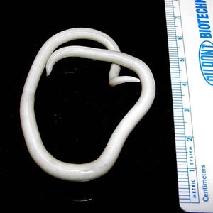
A. lumbricoides is characterized by its great size. Males are 2â€"4 mm in diameter and 15â€"31 cm long. The male's posterior end is curved ventrally and has a bluntly pointed tail. Females are 3â€"6 mm wide and 20â€"49 cm long. The vulva is located in the anterior end and accounts for about one-third of its body length. Uteri may contain up to 27 million eggs at a time, with 200,000 being laid per day. Fertilized eggs are oval to round in shape and are 45-75 μms long and 35-50 μm wide with a thick outer shell. Unfertilized eggs measure 88-94 μm long and 44 μm wide.
Epidemiology
_-Weisker-.jpg/800px-Modelle_von_Ascaris_lumbricoides_L._(Spulwurm)_-Weisker-.jpg)
More than 2 billion people are affected by this infection. The United States has a reported prevalence of 0.8% of the total population as of 1987. A. lumbricoides eggs are extremely resistant to strong chemicals, desiccation, and low temperatures. The eggs can remain viable in the soil for several months or even years.
Eggs of A. lumbricoides have been identified in archeological coprolites in the Americas, Europe, Africa, the Middle East, and New Zealand, the oldest ones being more than 24,000 years old.
Infections

Infections with these parasites are more common where sanitation is poor, and raw human feces are used as fertilizer.
Symptoms
Often, no symptoms are seen with an A. lumbricoides infection. However, in the case of a particularly bad infection, symptoms may include bloody sputum, cough, fever, abdominal discomfort, intestinal ulcer, passing worms, etc. Ascariasis is also the most common cause of Löffler's syndrome worldwide. Accompanying symptoms include pulmonary infiltration, eosinophilia, and radiographic opacities
Prevention
Preventing any fecal-borne disease requires educated hygienic habits/culture and effective fecal treatment systems. This is particularly important with A. lumbricoides because its eggs are one of the most difficult pathogens to kill (second only to prions), and the eggs commonly survive 1â€"3 years. A. lumbricoides lives in the intestine where it lays eggs. Infection occurs when the eggs, too small to be seen by the unaided eye, are eaten. The eggs may get onto vegetables when improperly processed human feces of infected people are used as fertilizer for food crops. Infection may occur when food is handled without removing or killing the eggs on the hands, clothes, hair, raw vegetables/fruit, or cooked food that is (re)infected by handlers, containers, etc. Bleach does not readily kill A. lumbricoides eggs, but it will remove their sticky film, to allow the eggs to be rinsed away. A. lumbricoides eggs can be reduced by hot composting methods, but to completely kill them may require rubbing alcohol, iodine, specialized chemicals, cooking heat, or "unusually" hot composting (for example, over 50°C (120°F) for 24 hours [1]).
Details of infection process
Infections happen when a human swallows water or food contaminated with unhatched eggs, which hatch into juveniles in the duodenum. They then penetrate the mucosa and submucosa and enter venules or lymphatics. Next, they pass through the right heart and into pulmonary circulation. They then break out of the capillaries and enter the air spaces. Acute tissue reaction occurs when several worms get lost during this migration and accumulate in other organs of the body. The juveniles migrate from the lung up the respiratory tract to the pharynx where they are swallowed. They begin producing eggs within 60â€"65 days of being swallowed. These are produced within the small intestine, where the juveniles mature. It might seem odd that the worms end up in the same place where they began. One hypothesis to account for this behavior is that the migration mimics an intermediate host, which would be required for juveniles of an ancestral form to develop to the third stage. Another possibility is that tissue migration enables faster growth and larger size, which increases reproductive capacity.
Diagnosis and treatment
,_Mina_de_sal_de_Hallstatt.JPG/323px-Cuc_intestinal_(Ascaris_lumbricoides),_Mina_de_sal_de_Hallstatt.JPG)
Most diagnoses are made by identifying the appearance of the worm or eggs in feces. Due to the large quantity of eggs laid, physicians can diagnose using only one or two fecal smears.
Infections can be treated with drugs called ascaricides. The treatment of choice is mebendazole. The drug functions by binding to tubulin in the worms' intestinal cells and body-wall muscles. Nitazoxanide and ivermectin can also be used.
References

External links

- Ascaris lumbricoides Video - DAVE Project
- Ascaris lumbricoides Poll - Research
- Ascaris lumbricoides image library