The uterine sarcomas form a group of malignant tumors that arises from the smooth muscle or connective tissue of the uterus.
Histology
_(2145766681).jpg/120px-Uterine_Tumor_in_a_33-Year-Old_(1)_(2145766681).jpg)
Tumoral entities include leiomyosarcomas, endometrial stromal sarcomas, carcinosarcomas and "other" sarcomas.
- If the lesion originates from the stroma of the uterine lining it is an endometrial stromal sarcoma.
- If the uterine muscle cell is the originator the tumor is a uterine leiomyosarcoma.
- Carcinosarcomas comprise both malignant epithelial and malignant sarcomatous components.
Classification

Leiomyosarcomas are now staged using the 2009 FIGO staging system (previously they were staged like endometrial carcinomas) at time of surgery.
- Stage I: tumor is limited to the uterus
- IA: ≤5 cm in greatest dimension
- IB: >5 cm
- Stage II: tumor extends beyond the uterus, but within the pelvis
- IIA: involves adnexa of uterus
- IIB: involves other pelvic tissues
- Stage III: tumor infiltrates abdominal tissues
- IIIA: 1 site
- IIIB: >1 site
- IIIC: regional lymph node metastasis
- Stage IVA: invades bladder or rectum
- Stage IVB: distant metastasis (including intraabdominal or inguinal lymph nodes; excluding adnexa, pelvic and abdominal tissues)
Endometrial stromal sarcomas and uterine adenosarcomas are classified as above, with the exception of different classifications for Stage I tumors.
- Stage I: tumor is limited to the uterus
- IA: limited to endometrium/endocervix
- IB: invades <½ myometrium
- IC: invades ≥½ myometrium
Finally, malignant mixed Müllerian tumors, a type of carcinosarcoma, are staged similarly to endometrial carcinomas.
- Stage I: tumor is limited to the uterus
- IA: invades <½ myometrium
- IB: invades ≥½ myometrium
- Stage II: invades cervical stroma, but no extension beyond the uterus
- Stage III: local and/or regional spread
- IIIA: invades uterine serosa and/or adnexa
- IIIB: vaginal and/or parametrial involvement
- IIIC: metastases to pelvic and/or paraaortic lymph nodes
- IIIC1: positive pelvic nodes
- IIIC2: positive para-aortic lymph nodes
- Stage IVA: invades bladder and/or bowel mucosa
- Stage IVB: distant metastases (including intra-abdominal metastases and/or inguinal lymph nodes)
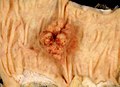
Signs and Symptoms
_(2145766681).jpg/800px-Uterine_Tumor_in_a_33-Year-Old_(1)_(2145766681).jpg)
Unusual or postmenopausal bleeding may be a sign of a malignancy including uterine sarcoma and needs to be investigated. Other signs include pelvic pain, pressure, and unusual discharge. A nonpregnant uterus that enlarges quickly is suspicious. However, none of the signs are specific. Specific screening test have not been developed; a Pap smear is a screening test for cervical cancer and not designed to detect uterine sarcoma.
Diagnosis
_(2145764413).jpg/800px-Uterine_Tumor_in_a_33-Year-Old_(2)_(2145764413).jpg)
Investigations by the physician include imaging (ultrasound, CAT scan, MRI) and, if possible, obtaining a tissue diagnosis by biopsy, hysteroscopy, or D&C. Ultimately the diagnosis is established by the histologic examination of the specimen. Typically malignant lesions have >10 mitosis per high power field. In contrast a uterine leiomyoma as a benign lesion would have < 5 mitosis per high power field.
Management
Therapy is based on staging and patient condition and utilizes one or more of the following approaches. Surgery is the mainstay of therapy if feasible involving total abdominal hysterectomy with bilateral salpingo-oophorectomy. Other approaches include radiation therapy, chemotherapy, and hormonal therapy.
Prognosis is relatively poor.
Epidemiology

Uterine sarcoma are rare, out of all malignancies of the uterine body only about 4% will be uterine sarcomas. Generally, the cause of the lesion is not known, however patients with a history of pelvic radiation are at higher risk. Most tumors occur after menopause. Women who take long-term tamoxifen are at higher risk.
