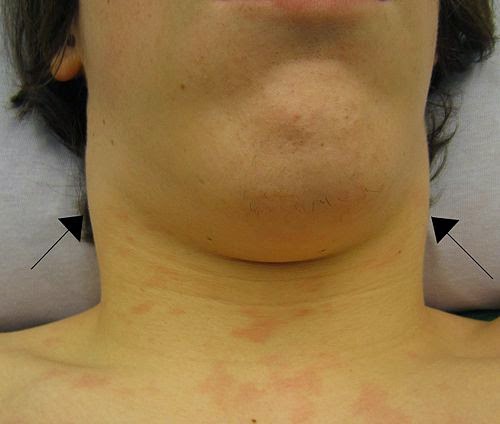
Lymphadenopathy refers to lymph nodes which are abnormal in size, number or consistency and is often used as a synonym for swollen or enlarged lymph nodes. Common causes of lymphadenopathy are infection, autoimmune disease, or malignancy.
Inflammation as a cause of lymph node enlargement is known as lymphadenitis. In practice, the distinction between lymphadenopathy and lymphadenitis is rarely made. Inflammation of the lymphatic vessels is also known as lymphangitis. Infectious lymphadenitides affecting lymph nodes in the neck are often called scrofula.
The term comes from the word lymph and a combination of the Greek words αδÎνας, adenas ("gland") and παθεία, patheia ("act of suffering" or "disease").
Types

- Localized lymphadenopathy: due to localized spot of infection e.g., an infected spot on the scalp will cause lymph nodes in the neck on that same side to swell up
- Generalized lymphadenopathy: due to a systemic infection of the body e.g., influenza or secondary syphilis
- Persistent generalized lymphadenopathy (PGL): persisting for a long time, possibly without an apparent cause
- Dermatopathic lymphadenopathy: lymphadenopathy associated with skin disease.
Causes

Lymph node enlargement is recognized as a common sign of infectious, autoimmune, or malignant disease. Examples may include:
- Reactive: acute infection (e.g., bacterial, or viral), or chronic infections (tuberculous lymphadenitis, cat-scratch disease).
- The most distinctive sign of bubonic plague is extreme swelling of one or more lymph nodes that bulge out of the skin as "buboes." The buboes often become necrotic and may even rupture.
- Infectious mononucleosis is an acute viral infection caused by Epstein-Barr virus and may be characterized by a marked enlargement of the cervical lymph nodes.
- It is also a sign of cutaneous anthrax and Human African trypanosomiasis
- Toxoplasmosis, a parasitic disease, gives a generalized lymphadenopathy (Piringer-Kuchinka lymphadenopathy).
- Plasma cell variant of Castleman's disease - associated with HHV-8 infection and HIV infection
- Mesenteric lymphadenitis after viral systemic infection (particularly in the GALT in the appendix) can commonly present like appendicitis.
Less common infectious causes of lymphadenopathy may include bacterial infections such as cat scratch disease, tularemia, brucellosis, or prevotella.
- Tumoral:
- Primary: Hodgkin lymphoma and non-Hodgkin lymphoma give lymphadenopathy in all or a few lymph nodes.
- Secondary: metastasis, Virchow's Node, neuroblastoma, and chronic lymphocytic leukemia.
- Autoimmune etiology: systemic lupus erythematosus and rheumatoid arthritis may have a generalized lymphadenopathy.
- Immunocompromised etiology: AIDS. Generalized lymphadenopathy is an early sign of infection with human immunodeficiency virus (HIV), the virus that causes acquired immunodeficiency syndrome (AIDS). "Lymphadenopathy syndrome" has been used to describe the first symptomatic stage of HIV progression, preceding a diagnosis of AIDS.
- Bites from certain venomous snakes such as the pit viper
- Unknown etiology: Kikuchi disease, progressive transformation of germinal centers, sarcoidosis, hyaline-vascular variant of Castleman's disease, Rosai-Dorfman disease, Kawasaki disease, Kimura disease
Benign (reactive) lymphadenopathy

Benign lymphadenopathy is a common biopsy finding, and may often be confused with malignant lymphoma. It may be separated into major morphologic patterns, each with its own differential diagnosis with certain types of lymphoma. Most cases of reactive follicular hyperplasia are easy to diagnose, but some cases may be confused with follicular lymphoma. There are six distinct patterns of benign lymphadenopathy:
- Follicular hyperplasia: This is the most common type of reactive lymphadenopathy.
- Paracortical hyperplasia/Interfollicular hyperplasia: It is seen in viral infections, skin diseases, and nonspecific reactions.
- Sinus histiocytosis: It is seen in lymph nodes draining limbs, inflammatory lesions, and malignancies.
- Nodal extensive necrosis
- Nodal granulomatous inflammation
- Nodal extensive fibrosis (Connective tissue framework)
- Nodal deposition of interstitial substance
These morphological patterns are never pure. Thus, reactive follicular hyperplasia can have a component of paracortical hyperplasia. However, this distinction is important for the differential diagnosis of the cause.
Localization

- Mediastinal lymphadenopathy
- Bilateral hilar lymphadenopathy
